
Why Binocular Vision and Depth Perception Matter for Ear Wax Removal with Dr Jan Rombout ENT
Ear Wax Removal: Clinical Insights with Dr Jan Rombout, Episode 2
In Episode 2 of Ear Wax Removal: Clinical Insights, ENT surgeon Dr Jan Rombout expands on one of his key factors and why he considers Binocular vision and Depth Perception so important when removing ear wax.
What is Depth Perception and why is it essential for Ear Wax Removal?
When performing ear wax removal, seeing the ear canal is only part of the challenge. The clinician also needs to understand where the instrument or microsuction tube is within the three-dimensional space of the ear canal.
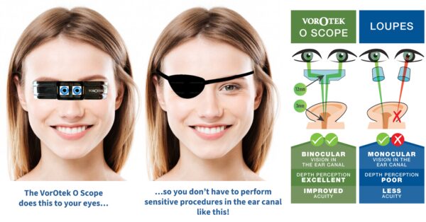
Direct binocular vision allows the brain to combine the slightly different images received by each eye, creating a three-dimensional perception of the clinical environment.
For ear wax removal, this means the clinician can more accurately judge the position of the instrument relative to the canal wall, wax and sensitive anatomy.
This becomes particularly important when working deeper within the ear canal, where precision and controlled instrument movement are essential.
Because direct binocular vision works with the body’s natural hand-eye coordination, the visual information and hand movement are closely connected. With appropriate training and experience, clinicians can develop an intuitive understanding of instrument position and quickly build confidence in safe and effective practice.
How can Clinicians optimise Depth Perception for Ear Wax Removal?
There are several technologies available to clinicians performing procedures within the ear canal, but as ENT Surgeon Jan Rombout further explains, they don’t all provide the same visual experience.
ENT fixed microscopes provide excellent magnification and depth perception. However, maintaining the optimal focal position can be restrictive during a procedure, especially if the patient or the microscope moves. They are also an expensive option for clinics conducting only simple ear assessments and procedures, like ear wax removal. Adding to this they tend to take up a large footprint in the clinic room, which for many clinics is prohibitive due to the lack of available space.
Surgical loupes provide magnification and portability, but conventional monocular viewing can limit the clinician’s visual information in a narrow space, due to the lack of depth perception.
Head-worn binocular systems provide another option. Like the ENT fixed microscope, there are specialised head-worn devices with converged binocular optics that provide stereoscopic vision directly in the clinician’s field of view. Combined with integrated illumination, this provides depth perception without requiring the clinician to work from a fixed viewing position.
The head-worn design also provides greater mobility and a wide practical working range, supporting efficient clinical and patient workflows.
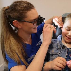
For ear wax removal, this combination is particularly relevant. Clinicians need sufficient visual detail while maintaining awareness of the instrument or microsuction tube within the three-dimensional space of the ear canal, but do not require the more complex settings of a fixed microscope.
The goal isn’t necessarily maximum magnification. It is sufficient magnification combined with direct binocular vision, illumination, accessibility and freedom of movement.
The right visualisation system helps the clinician see all of the ear canal clearly while allowing them to work naturally and intuitively.
When precision matters, the view matters.
You can watch clinical insights from Dr Jan Rombout in previous episodes via this link and enjoy more in the upcoming episodes of this series.
In the meantime you can follow these links for more information in relation to Direct Binocular Vision and Microsuction for cerumen management.